Behind every retinal disease is a clock counting down on photoreceptors – the light-sensing cells that once lost do not naturally grow back. A new study suggests that might change. Scientists show that instead of replacing lost cells, they can convince nearby cells in the eye to become them.

Image Source: https://www.pexels.com/photo/behind-blue-eyes-27616422/
The retina, tucked away at the back of the eye is a thin layer of tissue that works like a high-tech camera sensor. It contains neurons called photoreceptors which detect light and send them as visual information to the brain.
Failing to address retinal diseases such as retinal pigmentosa (genetic disorder causing photoreceptor degeneration), diabetic retinopathy (a diabetic complication caused by damage to retinal blood vessels), and age-related macular degeneration (damage to the centre of the retina called macula, responsible for high visual acuity), can result in an irreversible photoreceptor loss consequently leading to visual impairment. Such diseases currently have limited treatment options, hence finding new methods to regenerate retinal tissue is an overarching goal in ophthalmic care.
Previous attempts to rescue degrading retinas were done with cell-replacement strategies i.e. replacing injured cells with healthy ones, either using (i) stem cells – naive cells which can develop into any kind of cell, or (ii) progenitor cells from fetal retinas – immature cells from the foetus that can mature into specialized retinal cells. These cells had to go through lengthy protocols in order to be directed into retinal type. But these approaches are time-consuming and often showed inconsistencies in effectively replacing the photoreceptors. This happened mainly because it is difficult to produce a large volume of cells that would be required for sufficient replacement of damaged ones.
Direct Chemical Reprogramming:
As an alternative, a study by Yeuh-Chang Lee et al., preferred a ‘direct chemical reprogramming’ approach wherein there is no genetic manipulation or induction using external proteins/factors (which control genes by either turning them on or off in a cell). Instead cells are directly subjected to change from one form to another using a specific combination of chemicals.
Usually fibroblasts (fibrous type of cells that make up connective tissues) are the gold standard for cellular reprogramming procedures as they are easy to collect and grow abundantly in the lab.
In this case scientists exposed fibroblasts from human Tenon’s capsule (which supports and protects the eyeball), to a ‘chemical potion’ which is capable of altering signaling pathways inside cells. This can transform their identity into retinal progenitor-like cells, which are immature cells that later become functioning retina.
(When similar reprogramming conditions were applied to fibroblasts derived from other anatomical sources such as fetal lung fibroblasts and adult dermal fibroblasts, they were found to exhibit limited alteration. So, Tenon’s capsule was best suited for this study)
Basically, the process is a safe biochemical makeover rather than a complex DNA rewrite.
Methodology & Results:
Human Tenon’s capsule fibroblasts – HTFs, were excised from surgically discarded or donated ocular tissues and grown in the lab. Fibroblastic cells typically appeared adherent, flat and spindle-shaped under the microscope in cell culture dishes.
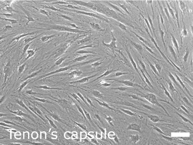
Fig 1: Flat, spindle-shaped fibroblast cells
(Image Source: Figure 1A from Yeuh-Chang Lee et al.
is licensed under Deed – Attribution 4.0 International – Creative Commons)
The researchers then administered a judiciously developed six-compound cocktail to the cells for over five days. One of the important factors was valproic acid (VPA) that helps loosen chromatin (a highly organized packing of DNA and proteins within the nucleus of the cell) and activate genes which can change the fate of cells from one type to another. Vitamin C was also used to support cell survival during the transformation.
The fibroblasts were observed to gradually transition from a flattened structure to dome-shaped with bright nuclei resembling retinal progenitor cells.
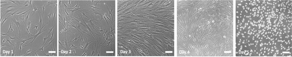
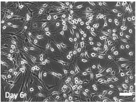
Fig 2: Day 1 to Day 6 transition of fibroblasts to dome-shaped retinal progenitor cells with bright nuclei
(Image Source: Figure 2E from Yeuh-Chang Lee et al.
is licensed under Deed – Attribution 4.0 International – Creative Commons)
More crucially, major changes in gene expression were noted. Genes associated with fibroblasts became less active, while genes related to retinal development became strongly activated, indicating that cells embraced their new identity.
Gene expression profiling confirmed several important retinal progenitor genes as a mark of successful reprogramming. One key marker was PAX6, a master control gene which regulates the formation of retinal tissue. Genes like VSX2, NEUROD1 and CRX which are involved in early retinal specification were also present. Additional markers included RHO and NRL, both responsible for photoreceptor development.
Further molecular analysis revealed that a substantial proportion of the induced cells expressed neural and retinal lineage associated proteins implying that changes had not only transcribed at the mRNA level but had also translated into protein expression.
Beyond gene expression and molecular characteristics, functional behavior was evaluated by monitoring calcium activity inside the cell. In-vitro (inside the cell culture dish) calcium imaging analysis proved that chemically induced cells displayed active calcium signaling when fed with glutamate (amino acid), whereas parental fibroblasts did not under the same experimental conditions. This aligns with the fact that neuronal populations in the retina like photoreceptors are known to use glutamate as their primary neurotransmitter for normal functioning.
For the final aspect of the study, researchers investigated whether the reprogrammed cells could survive after being injected into the retinal environments of lab rats.
Despite the high sensitivity and structural complexity of the eye, the transplanted retinal progenitor-like cells not only integrated and survived but also displayed recognizable retinal layering and detectable signs of functioning, as confirmed from imaging, histological (tissue) and electroretinogram (ERG – eye test that measures the electrical activity of the retina in response to light) analyses, one month after the transplantation.
Conclusions:
The research is still in its initial chapters. However, the results look hopeful thus far, prompting a lot more work that needs to be done before the method can be practically used in hospitals.
A number of limitations need to be addressed in future investigations:
First, a uniform conversion of cells from fibroblastic to retinal should be established using larger cohorts.
Second, more retina-specific functional assays have to be incorporated as recording calcium dynamics alone does not equate to complete physiological suitability.
Third, long term efficacy of in-vivo (animal) experiments must be assessed using extended follow-up and higher-resolution evaluations.
Broadly, the findings are both encouraging and impactful, offering tremendous promise for restoring sight and therefore improving the quality of life.
Edited by Olivia Fish & JP Flores


Leave a comment