Optimal eye placement across the animal kingdom is a diverse phenomenon to suit every creature’s purpose and way of life. During development, the interocular distance (space between two eyes) and eye dimensions are dictated by master genes that define the early eye field.

Image from Pexels
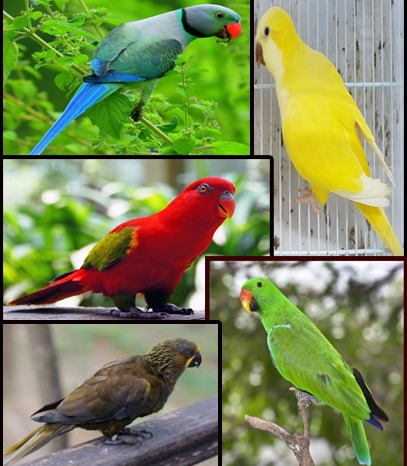
From the wide-set eyes of a hammerhead shark to the front-facing gaze of an owl, some animals sport eyes on the sides of their heads for panoramic vision, whereas others have them front and center for focus and depth.
These positions and sizes are orchestrated by cell signaling molecules called ‘morphogens’, which help shape the tissues and organs in a developing embryo. Cells respond to different levels of morphogens by activating specific genes, which guide their fate in the body.
Wingless (Wg), which belongs to the Wnt family of signaling proteins, is one such molecule important for craniofacial (face and skull) development, especially in the positioning of the eyes in the head.
But how exactly is Wg regulated to produce ‘eyes’ of the right size that get placed at the right spot on the head?
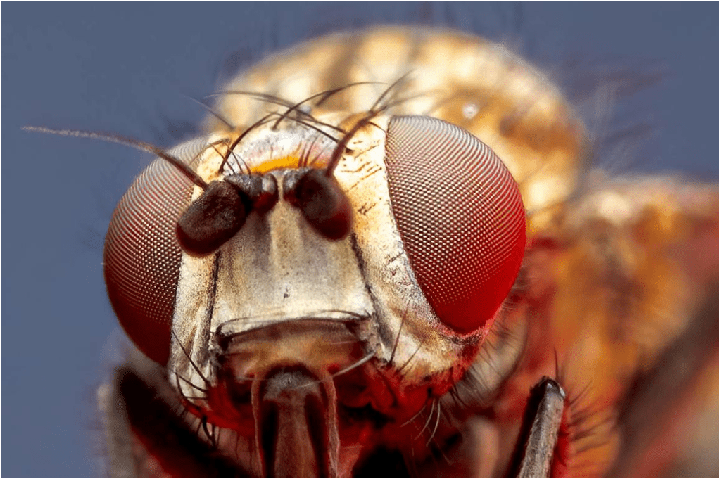
In a 2024 PNAS study, researchers from the USA and Japan conjointly elucidated a critical role for a particular gene called the Defective proventriculus (Dve) in fruit flies (Drosophila), which acts like a master switch controlling the levels of Wg in the developing eye-antennal disc.
Dve is expressed in the dorsal head vertex region– the uppermost back of the head.
By regulating Wg, Dve ensures that an apt number of cells become a part of the eyes, thus distinctly demarcating the eyes in the head space. If Dve is shunned, then too many cells might become eyes, leading to problems like magnified eyes or abnormal eye placements.
After running through a series of experiments at the molecular level, researchers underscored that a Loss-of-function (LOF) of Dve results in eye enlargements indicating an expanded eye field, whereas a Gain-of-function (GOF) of Dve leads to reduced eye size and suppression of retinal determination genes such as Eya and Dac, while the general eye specification gene Ey remains unaffected. This implies that Dve is indispensable for retinal development.
On the other hand, Wg inhibition in Dve expressing cells causes the entire head to transform into eyes, thereby highlighting the significance of a properly functioning Dve-Wg cascade in maintaining the appropriate ratio of eye-versus-head fate.
This finding represents an important potential for translation to human biology. By the same token, the human ortholog SATB1 exhibits similar functions in regulating Wg signaling. Dysregulation of SATB1 can lead to craniofacial abnormalities like hypertelorism (increased distance between the eye orbits) and dystopia (distorted placement of eyes). There might be an evolutionarily conserved mechanism between Dve and SATB1 in eye patterning that can tell us more about human eye biology.
There are a variety of complex signaling interactions that take place during eye growth from the embryonic stage that pave the way for deeper analyses of other genes that may come into the picture. In the long term, these findings could even open up potential therapeutic targets for related birth defects.
After all, there is always much more than what meets the eye!
Edited by Aanchal Saxena & Jayati Sharma


Leave a comment